


CX NEWS
Early results from subclavian artery branched endoprosthesis studies presented at CX
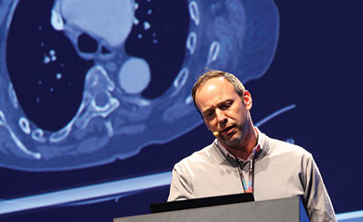
Michael Dake, Stanford, USA, outlined the first experiences with the Gore TAG (W L Gore) thoracic branch endoprosthesis and Frank R Arko, Charlotte, USA, presented on the MONA LSA (Medtronic) branched device results at a session yesterday. Both speakers shared early cases and their results.
Thoracic aortic aneurysms that involve the left subclavian artery often leave physicians no choice but to use surgical techniques or to cover the branch vessel. “Although reports from single-centre experience with the hybrid approach have been positive, a single branch thoracic endograft specifically designed for treatment of the aortic arch (Zone 0–2) could be useful in extending the advantages of endovascular repair to the aortic arch. As such, the Gore TAG Thoracic Branch Endoprosthesis is designed as a modular system which allows for treatment of aortic arch pathologies using a less invasive hybrid endovascular approach,” Dake said.
He presented on the Zone 2 US investigational device exemption feasibility trial that will enrol 20 to 40 patients at six sites. Patients will be followed for five years. The study will evaluate the device for the treatment zone 2 aneurysms.
The Gore TAG thoracic branch endoprosthesis has a modular construction design with off-the-shelf components. It has an inner lumen for anchoring and sealing the branch component. The complete system consists of aortic and branch components designed for the use in the arch, and also the accompanying accessory devices to facilitate delivery.
“The system is easy to use with a single femoral access and requires minimal catheter manoeuvres. It is safe with zero ischaemic time and has low risk of branch vessel coverage. After the first four cases, successful access and deployment of the endoprosthesis was seen in all cases. There was one procedural type 1 endoleak that resolved without re-intervention at one month. There were no device-related endoleaks, but one type 2 endoleak was seen at one month. There were no deaths or neurological events. There were no site reported serious adverse events related to the device,” he said.
Dake explained that the procedure to deploy the device included inserting the guidewires in aorta and branch vessel; introducing the aortic component over both guidewires into position within the arch; deploying aortic component and withdrawing catheter; advancing the introducer sheath and dilator and advancing and deploying branch component.
In order to be enrolled, patients had to have descending thoracic aortic aneurysms requiring placement of the proximal extent of the aortic stent graft in Zone 2. The primary endpoints of the study were: successful access and deployment of the thoracic branched endograft and the primary patency of the side branch assessed by angiography when the procedure finishes. The secondary endpoints included a one-month core lab analysis, assessing the side branch primary patency and device-related endoleaks.
Arko told delegates that there was a clinical need for left subclavian artery preservation in association with encroaching thoracic artery aneurysm. He presented the current status of an early feasibility trial in the FDA’s new innovation pathway testing the Valiant Mona LSA device.
The key goals of the trial are to validate the procedure in humans; assess the safety and performance acutely and at 30 days and collect imaging data to augment the current understanding.
Arko told delegates that the Valiant Mona LSA Thoracic Stent Graft consisted of a flexible cuff “volcano” on the main body. The system is a two-graft system and the main graft system comes in diameters between 30 and 46mm and in the single length of 15cm. It is a two-wire system as well and the secondary wire can be snared and then the second branch is brought up and deployed. The branch graft itself is made of a nitinol helical wire and polyester fabric. It has a proximal flare. The branch graft is always 40mm in length and a 15F profile, femoral access system.
The early feasibility trial is a prospective, non-randomised, three-centre, premarket clinical study that has enrolled nine patients. The primary safety and effectiveness objectives were measured acutely and at 30 days. “The follow-up schedule will be at 0-30 days, six months, 12 months and annually through five years,” Arko said.
The principal investigators are Eric E Roselli, Cleveland, USA, Frank R Arko, Charlotte, USA and Matt Thompson, London, UK.
Arko revealed that the current status of the early feasibility trial had seven patients enrolled as part of the US cohort and two enrolled as part of the cohort outside the US. One emergent case had been enrolled in the USA, outside of trial. Acute procedure results from all seven patients revealed 100% technical success and 100% patency in both main and branch stent graft. There were no type I or III endoleaks.
“All current devices need a healthy landing zone to seal. To achieve sufficient landing zone, the left subclavian artery may be sacrificed with resulting complications, such as 6% arm ischaemia, 4% spinal cord ischaemia, 2% vertebrobasilar ischaemia, 5% anterior circulation stroke and 6% death. Left subclavian artery preservation is recommended in the literature. 17–43% of patients undergoing TEVAR have planned coverage of the left subclavian artery to achieve an adequate proximal seal and the coverage of the left subclavian artery without revascularisation is the single most important predictor of post-TEVAR stroke,” Arko said.
CX ilegx Collaboration Day hosts Endovascular Electronic Education and focuses on key approaches to save limbs
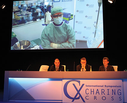
At the ilegx Collaboration Day, attendees learnt about the best management therapies for diabetic foot care and vascular reconstruction in critical limb ischaemia patients and for the second year running, the symposium broadcast edited live cases treating superficial femoral artery lesions to the Far East and North America in an event named Endovascular Electronic Education, sponsored by Abbott Vascular.
In the morning, three edited live cases were showcased to the Far East. The first case was performed by Andrej Schmidt in Leipzig, Germany, the second procedure was carried out by Peter Goverde in Antwerp, Belgium, and the third case was performed by Josef Tacke in Passau, Germany. The Abbott Supera stent system was used to treat occlusion and total occlusion of the superficial femoral artery and proximal popliteal artery.
Schmidt, Goverde and Tacke were present to answer questions from the audience at CX and those watching the live broadcast in the Far East.
Responding to questions about how a physician chooses a stent, Schmidt said: “In some cases it is clear—if it is a calcified lesion, for me it is clear that a drug-eluting balloon might not be so good, we know this from our data, and the Supera stent is the stent of choice.” He added that more data are required to make the stent selection process more comprehensive.
In the afternoon, edited live cases were broadcast to North America.
Early referral, fast track care and multidisciplinary work: key approach to save limbs
The ilegx initiative, launched in 2008, was created in response to the increasing number of lower limb amputations which are mostly due to type II diabetes. Michael Edmonds (London, UK) who is one of the founders of ilegx, introduced the “diabetic foot care” session, which was organised in conjunction with the King’s College Hospital Open Access System, with a presentation which highlighted the importance of early diabetic foot referral and interdisciplinary work as an effective approach to reduce the number of lower limb amputations, flagship principles of the ilegx initiative. He said: “Up until recently, the diabetic ischaemic foot has defeated every health care system in the world. However, a strategy which combines early referral and interdisciplinary working has led to improvements in care.” With this in mind, he commented, “ulcers can now be healed and amputations can be prevented.”
Edmonds also referred to the importance of organising a “fast-track” service in a “one-stop” visit, comprising clinical assessment, same-day investigations and urgent management to treat infection and revascularise the foot, when dealing with cases of “diabetic foot attack.” “This is best carried out in a diabetic foot clinic which can see the ischaemic patient in an open access system without delay and has rapid availability of debridement and intravenous antibiotics to treat infection and control the septic vasculitis.” He also said that diabetic foot patients who experience repeated crises from the rapid onset of infection need a special form of such easily accessible care provided by the diabetic foot clinic. “Such integrated fast track care can rapidly restore the circulation, limit tissue necrosis, save the limb from amputation and provide complete recovery from the foot attack,” he commented.
An interdisciplinary team including podiatrists, nurses, orthotists, microbiologists, interventional radiologists and surgeons—including vascular surgeons, orthopaedic surgeons and plastic surgeons—is the ideal team required in the management of diabetic foot, Edmonds noted.
He also mentioned that to complete an effective management of diabetic foot, follow-up and rehabilitation are required.
Interdisciplinary views on diabetic foot care
A vascular surgeon, an interventional radiologist and a podiatrist from King’s College Hospital, London, UK, shared with CX delegates their best practices treating diabetic foot.
Vascular surgeon Hisham Rashid, said that “the incidence of type II diabetes mellitus is increasing across the world with an expected rise in 2030 to more than 12% in a large population of the world.”
This is a worrying figure taking into account that “a major amputation rate is significantly higher in diabetic patients,” he noted.
Rashid told CX delegates that aggressive revascularisation with angioplasty, bypass or hybrid techniques is essential for limb salvage and reduction of major amputation rate. He mentioned that at King’s College Hospital, 77% of cases are treated with angioplasty and 23% with distal bypass. He said that distal bypass surgery “plays a major role in revascularisation, especially in patients presenting with significant tissue loss and when angioplasty is not feasible.” Hybrid techniques in “no-option” critical leg ischaemia have also proven very effective in preventing major amputation in this challenging group of patients, he commented.
At King’s, Rashid noted, revascularisation using distal and ultra-distal bypass has a very good outcome with a one-year amputation rate of 3.4% and 30-days mortality rate of 1.5–1.7%. At one-year, “mortality is significantly influenced by end-stage renal failure and age rather than diabetes mellitus,” he said.
Interventional radiologist Dean Huang, said that the concept of “foot attack” and “time is tissue” in diabetic patients means that treatment of an infected ulcer should be handled as an emergency with the management of a multidisciplinary team. At King’s College Hospital, “we follow this approach; we work on the basis of rapid access to diagnosis, rapid access to intervention and follow-up of interventional and surgical procedures.” From the interventional radiologist perspective, he said, “rapid access to imaging diagnostics enables the selection and planning of the optimal strategy.” Ultrasound, CTA and MRA have their place and angiography remains the gold standard, he commented. “Prompt definitive treatment with radiological intervention and/or surgical bypass to revascularisation for healing in conjunction with wound care and antibiotics is the key to achieve prevention of amputation,” Huang noted. “The threshold of what can be treated with endovascular procedures is shifting as more sophisticated devices appear on the market.”
Podiatrist Jennifer Tremlet, spoke about the different techniques used at King’s College Hospital to heal diabetic foot wounds. “Diabetic foot patients are complex cases, they experience extensive tissue loss and infection. In order to achieve successful wound healing, they require intensive wound care and rigorous monitoring.” She said that, depending on the complexity of the wound and the type of patient, they use different multi-modal techniques including debridement, larvae therapy, hydrosurgery therapy topical negative pressure therapy, split skin grafts and pressure relief.
Foot care and arterial reconstruction
At a session on foot care and arterial reconstruction, Christopher Attinger (Washington, USA) said that using the angiosome principle to guide revascularisation is a critical component for optimising wound healing. He explained that the angiosome concept divides anatomic regions into three-dimensional blocks of tissue fed by source arteries and creates a framework for understanding tissue perfusion, predicting wound healing, and planning surgical interventions. Therefore, “understanding the boundaries of an angiosome and the vascular connections between source arteries provides the basis for limb salvage to optimise revascularisation to ischaemic areas and promote wound healing.”
Mauro Gargiulo (Bologna, Italy) spoke on the need to have multidisciplinary guidelines to support the treatment of critical limb ischaemia. He referred to a consensus document that has been recently published on the treatment of peripheral arterial disease in diabetes written by the Italian Societies of Diabetes (SID, AMD), Radiology (SIRM) and Vascular Endovascular Surgery (SICVE). The consensus, published in Nutrition, Metabolism & Cardiovascular Disease (Aiello A et al, 2014; 24: 355–369), higlights that “the prevalence of peripheral arterial disease is high in diabetic patients and, associated or not with peripheral neuropathy, can be found in 50% of cases of diabetic foot.” Gargiulo said that the document summarises indications for revascularisation, revascularisation techniques and details on follow-up of revascularised patients, among other topics.
In the same session, Bijan Modarai spoke about effective cell therapies for revascularisation of critical limb ischaemia and Roberto Ferraresi discussed patient-centric revascularisation strategies.
At the end of the ilegx Collaboration Day a roundtable consensus on the role of drug-eluting balloons for the treatment of superficial femoral artery including data from the IN.PACT SFA, ILLUMINATE, Levant 2 and BIOLUX-PI studies was held. Vascular News will be reporting on the outcomes of this discussion.
Physicians share new product ideas at “Speed Dating” event
For the first time at the Charing Cross Symposium, physicians shared their product ideas with experienced physician-inventors, engineers and marketing experts in an event called “Speed Dating”. The event was held at the Innovation Showcase on Sunday 6 April. Physician-inventor David King (London, UK) won the “Dragons’ Den CX Innovation Showcase prize”.
Jean Bismuth, Elika Kashef and Stephen Greenhalgh, chairs of the session, designed the event to provide a platform for physicians who are seeking advice from experts on how to put their business and product ideas onto market.
Bismuth explained the rationale for the event: “Many young physicians and even senior physicians—as they gain experience through the years—have a desire to design their own medical tools and they start thinking and developing the idea. Unfortunately, sometimes they cannot get their innovations onto the market because they do not have the access to the experts to take the idea to the next level. With this “Speed Dating” event, we want to collaborate to bridge that gap.”
At the event, physicians had the opportunity to speak first to a group of physicians who have experience developing devices, then to engineers and finally to marketing experts. “Protect your idea” was the main advice that experts gave to the participants.
After the “Speed Dating” event, the Dragons’ Den CX Innovation session took place and awarded David King with £1,000 for his innovative idea. King designed a Doppler device called “Blue Dop” that works via a tablet device. He said that it uses a special algorithm that can measure blood pressure without the need to use a cuff or needles. He noted that the device is unique in the market and can be used in renal dialysis, arterial disease and sports medicine.
CX exposes alarming lack of physician awareness about radiation hazards
A session held yesterday at the Charing Cross International Symposium revealed that there is a widespread lack of physician awareness about the dangers of radiation in the interventional suite. The effect of exposure to radiation has become particularly important with the move from open to endovascular procedures, and delegates heard how no single specialty had a monopoly on bad practice when it came to radiation.
During the session it became apparent that the knowledge base about radiation had not been developed and incorporated into training and that appropriate behaviour in the interventional suite was often ignored, leading to operators receiving unnecessary doses of radiation. Surgeons, who have belatedly started using radiation, interventional cardiologists, interventional radiologists and radiologists are all guilty of bad practice, delegates heard.
“We are not aware of how badly affected we can be and this session turns the spotlight on how little we know. It is very important to become aware and realise the danger before you can start steps to reduce it. Perhaps as a profession we are not doing enough,” said Roger Greenhalgh, London, UK, chairman of the symposium.
Lindsay Machan, Vancouver, Canada, who was invited to comment on the Radiation Exposure session, told delegates that there were “two dirty little secrets” about radiation that he wanted delegates to pay attention to. “The first is that there is no safe dose of radiation; the idea that there is a threshold has now been debunked. The second is that every person in this room has a variant level of the radiation repair genes and there is no test as yet for the repair genes.”
Machan referred to the results of a long-term international study of thousands of workers exposed to radiation in the Chernobyl disaster in 1986, which found that the disaster clean-up crew, no matter where they were located, had the same incidence of cataracts, dispelling the idea of there being a threshold for cataract development. Referring to the outcomes from another 20-year study published by Chodick et al in the American Journal of Epidemiology in 2008 that found that there was no apparent threshold level for technologists to get cataracts, he said: “Everything that you hear about thresholds is absolutely not true. Treat radiation like iodinated contrast and use only as much as needed and no more.”
Machan also told delegates about the work of the late Basil V Worgul, New York, USA, which had shown that there was a possibility that the human population included genetically predisposed radiosensitive subsets.
“Everybody in this room has some degree of radiation damage and not everyone has complex innovations available to help them reduce the dosage. However, the distance from the tube and the importance of magnification cannot be overstated,” Machan said.
A member of the audience referred to currently available tools that enabled the visualisation of dosage in the form of a light or sound alarm. “Everybody wants to avoid radiation, they are just not aware of how their behaviour impacts dose. So if you stand unnecessarily close to the machine, there is an immediate warning and this can teach operators a lot,” he said.
Operator behaviour is driven by various factors
“You cannot overstate the importance of a real time reminder of the dose. Another important thing we have observed is that when our nurses and technologists became aware of their own risk, it changed the dynamic quite considerably. Operators are driven by a different dynamic; we want to get that procedure done, we want to show the photos and have a tendency to ignore [the radiation dose] and the consequences as they might be 20 years away. However, the nurses and technologists are not [driven by the same things]. They are there to do a job. As soon as they realise that they are at risk, it has an amazing impact. They start pointing out the fluoro time, question where they are asked to stand, and if you are doing a pedal puncture they might not stand behind the foot and hold the foot for you,” Machan said.
Another member of the audience made the point that they had measured and found that the radiation exposure was less in the hybrid suite as compared to their previous set-up. Their team had also found that the operator steps away just 6% of the time during digital subtraction angiography turns and have been working on educating the entire team. The awareness is lamentable, delegates heard.
Patients suffering burns
“One of the problems is that radiation burns can occur several months after the procedure and often the patient does not connect that to the procedure. Also, due to the fact that the image intensifier is above them, they think that the radiation dose is there. They do not realise why they have a burn on their back and often go to see a physician who does not have radiation in their mind as the cause, so there is a gross underreporting of the problem,” Machan said. The panel also commented that the biggest risks were for health professionals, as they are exposed repetitively during the course of their work. The maximum exposure resulted when branched devices were implanted, and the biggest risks are for health professionals as they are exposed every day.
Is regulation around the corner?
Machan made the point that while the health effects of radiation were one aspect, another issue was that of looming regulation for radiation workers. He pointed to the current practice in some US hospitals of radiation workers “sitting it out” if they had a high reading on their dosimeters. The sit-out period could range from anywhere between a week and a month. “Think of what this would do to your endovascular practice,” urged Machan. “If you have to sit out for a month every couple of months, you cannot actually practise.”
The panellists noted that while there was a wide variation in education and training requirements for personnel to be able to use radiation, there was need for special education for the whole team on to behave in an operating room. Machan also noted that while there were well laid out and appropriate guidelines in place, that the bigger problem was with adherence and enforcement.
Johannes Gahlen, Ludwigsburg, Germany, who spoke on the importance of radiation dosage exposure to patient and operator, said: “It is vital to adhere to the ALARA principle of keeping the dose as low as reasonably achievable. It is important to decrease the beam time, keep your distance from the system, use protective shielding, instruct the team, collimate the field of view, reduce the frame rate use use low dose programmes,” he said.
Following Gahlen’s presentation, Koning spoke about the novel X-ray system and how it can reduce radiation doses for patient and staff during endovascular procedures. He presented the results of a study and said that initial findings indicate that a significant radiation dose reduction of up to 75% to staff and patient can be realised for specific procedures.
He continued saying that radiation reduction in complex procedures, such as fenestrated stent graft implantation, means that implementation of lower doses of radiation is sometimes not attainable. He said that reducing radiation dose during the procedure will typically result in lower image quality, which may be unacceptable for clinical success of the endovascular treatment.
In the study, the investigators upgraded their existing C-arch to AlluraClarity (Philips Healthcare), which reportedly resulted in an ability to lower the required radiation exposure without losing image quality and without changing the workflow of the physician.
Koning reported that the results of the study are currently being processed for scientific publication. However, he said that initial findings indicate a significant dose reduction can be achieved. He said image quality was judged to be equal or superior compared to the image quality before installation and interventional work flows were uninterrupted.
Concluding, Koning said: “With the latest generation in imaging systems, we also see promise in increasing patient and staff safety without sacrificing the imaging quality required to provide optimal care.”
Still a lot to learn about abdominal compartment syndrome
At CX 2014, a mini symposium on abdominal compartment syndrome shed light on how little is known about the condition. It became apparent that the syndrome still has not been clearly defined, many of its triggering factors have yet to be recognised, and its management is limited to surgical decompression. Therefore, preventing both intra-abdominal hypertension and preventing intra-abdominal aortic hypertension from developing into abdominal aortic compartment syndrome is key.
Jan de Waele (Ghent, Belgium) stated that abdominal compartment syndrome has gone from being a syndrome that many vascular surgeons thought is “imaginary” to one that was now “being taken seriously.” He added that, according to the definitions of the World Society of the Abdominal Compartment Syndrome (WASCS), the syndrome was now defined as sustained intra-abdominal pressure of 20mmHg and it is associated with new organ dysfunction. “It can be a combination of haemodynamic problems, respiratory dysfunction, metabolic acidosis, and acute kidney dysfunction,” de Waele commented. However, he reported that “organ dysfunction sets in if you just look for it” at the threshold for intra-abdominal hypertension (a sustained pressure of 12mmHg or more). Therefore, he said it “made sense” to take steps to prevent both intra-abdominal hypertension and abdominal compartment syndrome. De Waele added: “We know the patients who are at risk, so we can really target intervention and we a have an indicator available—the intra-abdominal pressure.”
Anders Wanhainen (Uppsala, Sweden) explained that the WASCS had identified that risk factors for intra-abdominal hypertension and abdominal compartment syndrome included diminished abdominal wall compliance, increased intra-abdominal contents, capillary leak/fluid resuscitation, and increased intra-luminal contents. He noted that many of these risk factors were “relevant for vascular surgeons, particularly those managing patients with ruptured abdominal aortic aneurysms.” For example, Wanhainen explained, ruptured aortic aneurysms are associated with major trauma, intra-abdominal fluid collection, acidosis, hypothermia, polytransfusion, and shock or hypotension. He added: “Repair of a ruptured abdominal aortic aneurysm is definitely a high-risk procedure for the development of abdominal compartment syndrome.” According to Wanhainen, a study found that about 50% of patients undergoing open repair develop intra-abdominal hypertension and about 20% develop abdominal compartment syndrome. He added that these figures were, respectively, about 20% and 10% in patients undergoing endovascular aortic aneurysm repair (EVAR) but commented: “The observed lower risk after EVAR for a ruptured abdominal aortic aneurysm will probably change when more patients in shock are treated with EVAR.”
Wanhainen stated that because of the risk of intra-abdominal hypertension and abdominal compartment syndrome, intra-abdominal pressure “can and should be measured, preferably at the bedside in the intensive care unit” in patients undergoing emergency surgery or EVAR for a ruptured abdominal aortic aneurysm. He added that it was “completely possible” to eliminate the problem of abdominal compartment syndrome by taking adequate preventative measures—these measured included early bleeding control (which he called “crucial”), judicious fluid resuscitation, and prophylactic open abdomen management in selected patients. He added that preoperative and intraoperative factors could help to determine which patients should be selected for open abdomen management.
Martin Björck (Uppsala, Sweden) also stated that prevention was important in the management of intra-abdominal hypertension/abdominal compartment syndrome. In the context of EVAR/open repair, he said that prevention “started at the operating table.” He noted that this involved “a massive transfusion protocol and avoiding crystalloids,” agreeing with de Waele that it was “crucial” to monitor intra-abdominal pressure in all patients after aortic aneurysm repair in the postoperative period.
Björck noted: “We prevent abdominal compartment syndrome with aggressive medical management [in patients with intra-abdominal hypertension] but sometimes abdominal decompression is necessary.” He added, in terms of medical management, early pain relief could be “remarkably effective” and neuromuscular blockade, if the patient needs mechanical ventilation, was also very effective.
Björck reported that the recently updated 2013 WASCS guidelines were able to give strong recommendations for the management of abdominal compartment syndrome. He said that the guidelines recommend decompression laparotomy if abdominal compartment syndrome was present, protocolled effort should be made to obtain an early abdominal closure as “severe complications” can occur if open abdomen therapy is prolonged, and strategies using negative pressure wound therapy should be used. Björck agreed with de Waele that open abdomen treatment as a preventative treatment was an option as it “made sense” not to close a tense abdomen [in patients undergoing open repair] even though there are no data to support this approach.
However, he added if delegates used the preventative open abdomen therapy, they should close the abdomen “quickly”.
De Waele also spoke about the management of intra-abdominal hypertension/abdominal compartment syndrome, focusing on critical care. He said that the introduction of medical management options to decrease intra-abdominal pressure had “significantly changed the management of patients with intra-abdominal hypertension” and expanded on the WASCS recommendations—“The WASCS medical management algorithm identified five targets for medical interventions such as nasogastric decompression, neuromuscular blocking agents and percutaneous drainage among others. It is estimated that medical management can avoid surgery in a large proportion of patients with impending abdominal compartment syndrome.” However, de Waele said that surgical decompression remained an important element in the armamentarium and “may still be required” in some patients with therapy resistant abdominal compartment syndrome and significant organ derangement.” Concluding, he said that the management of abdominal compartment syndrome had now become the management of intra-abdominal hypertension as monitoring intra-aortic pressure was the “first and essential step” and prevention should be used where possible.
Charing Cross chairman Roger Greenhalgh (London, UK) told CX Daily News: “If I were a young physician, looking for an area to elucidate, I would choose abdominal compartment syndrome.”
No type I and III endoleaks with the Incraft system for EVAR at two years
Giovanni Pratesi (Florence, Italy) reported that the two-year results of the INNOVATION study (A multicentre, open-label, prospective, non-randomised study of the Cordis Incraft system in subjects with abdominal aortic aneurysms) confirm the “very promising results” of the one-year outcomes of the Incraft bifurcated stent graft system (Cordis). The results, presented at the Charing Cross Symposium 2014 (5-8 April, London, UK), showed that the system was not associated with any endoleaks at two years in patients undergoing endovascular aortic aneurysm repair (EVAR).
Pratesi stated that the Incraft system - showcased for the first time at the Charing Cross Symposium - has been designed to overcome the limitations of current stent grafts for the management of abdominal aortic aneurysms. He commented that it has an ultra-low profile delivery system and had the ability to be “customised during the procedure thanks to the bilateral in-situ length adjustment features”. Also, according to Pratesi, the graft offers the possibility of covering a wide range of anatomies with a minimum number of product codes.
The aim of the INNOVATION study was to assess the safety and efficacy of the Incraft system for the management of aortic abdominal aneurysms. The primary endpoint was technical success at one-month and the one-year safety endpoints included the absence of device- or procedure-related major adverse events, absence of type I or III endoleaks and maintenance of device integrity through one-year of follow-up. The main inclusion criteria included a proximal neck length of ≥15mm and up to 27mm in diameter, an access vessel large enough to accept the 14F outer diameter of the delivery catheter, and an aortic bifurcation >18mm in diameter.
The rate of technical success at one month was 97% (56 of 58 patients of the original 60 patients who were enrolled) and the rate of freedom from aneurysm enlargement was 100% at one year with the absence of both type I and III endoleaks in all patients.
Pratesti reported that two-year results confirmed these “very promising” early and one-year outcomes. He added that (at two years): “There were zero incidences of endoleaks (type I or type III), device- or procedure-related major adverse events, stent-graft migrations, or stent fractures.” He further said that there were also no incidences of aneurysm sac enlargement with sac shrinkage being observed in 45% of patients. However, one patient did develop a late limb ischaemia occlusion at day 666 due to sac contraction and limb confirmation change.
Pratesi concluded: “These excellent results are encouraging and suggest that the Cordis abdominal aortic aneurysm stent graft is a valuable alternative device with increased applicability over the broad spectrum of aorto-iliac anatomic configurations encountered in patients undergoing EVAR.”